45 labeling gel electrophoresis
ImageJ for Editing & Labelling PCR Gel Image | Biotechnology This Tutorial is all about how to quickly Edit & Label PCR Gel Image Using ImageJ software. Presented by - Elvis SamuelJoin Our Telegram Channel for free Sof... Gel Electrophoresis Overview | Science Primer Place the gel in the gel box making sure the gel is completely submerged in the buffer and that the wells are oriented properly (closest to the negative, usually black, electrode). Add loading dye to the samples and standards. Pipette a small volume * of sample/standard into each well.
The in vivo isotopic labeling of proteins for polyacrylamide gel ... The in vivo isotopic labeling of proteins for polyacrylamide gel electrophoresis Methods Mol Biol. 1984;1:75-80. doi: 10.1385/-89603-062-8:75. Author J W Pollard 1 Affiliation ... wand cheap means of quantifying proteins separated by gel electrophoresis. ...

Labeling gel electrophoresis
Gel Electrophoresis - an overview | ScienceDirect Topics Gel electrophoresis is an analytical technique that allows size separation of DNA as well as other macromolecules. For gel electrophoresis, a DNA sample is loaded at one end of a gel matrix (usually agarose or acrylamide) that provides a uniform pore size through which the DNA molecules can move. Gel Shift Assays (EMSA) | Thermo Fisher Scientific - US Gel shift assays need not be limited to protein-DNA interactions. Protein-RNA and protein-peptide interactions have also been studied using the same electrophoretic principle. Overview of the gel shift assay method. The gel shift assay consists of three key steps: (1) binding reactions, (2) electrophoresis, (3) probe detection. Part 2: Analysing and Interpreting (Agarose) Gel Electrophoresis Results The agarose gel electrophoresis is a molecular genetic technique used to separate DNA on the basis of size and charge of it. The negatively charged DNA migrates towards the positive node under the influence of the current. The results of agarose electrophoresis are affected by some of the factors enlisted below, The concentration of gel
Labeling gel electrophoresis. PDF Gel Electrophoresis Size Marker - dia-m.ru Labeling in four positions via the terminal EcoR I generated recessed ends is possible, especially with the DNA ladder 100 bp (A3470) and the DNA Ladder Mix 100 - 5000 (A3660). ... The different gel formats for agarose and polyacrylamide gel electrophoresis and the varying sensitivity of staining or detec- Agarose gel electrophoresis of labeled DNA in which the same gel is ... Agarose gel electrophoresis of labeled DNA in which the same gel is displayed before staining (Unstained) and after ethidium bromide staining (EtBr Stained). Lanes C contain unlabeled pCI Luc... Two-Dimensional Gel Electrophoresis and 2D-DIGE - PubMed in particular, a modified version of 2d-page, two-dimensional difference gel electrophoresis (2d-dige), which uses differential labeling of protein samples with up to three fluorescent tags, offers greater sensitivity and reproducibility over conventional 2d-page gels for differential quantitative analysis of protein expression between … PDF Disposal of Electrophoresis Gels and Solutions - Cornell University Utilize the following procedures for each specific type of electrophoresis gel waste. ELECTROPHORESIS GELS AND CONTAMINATED "NON-SHARP" LAB DEBRIS . 1. COLLECT: Collect electrophoresis gels and contaminated "nonsharp" lab debris (e.g., gloves, pads, towels, - tubes, etc.) into a 5gallon pail lined with a clear plastic - liner. The 5 ...
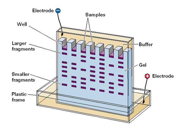
PDF Protocol 4: Gel Electrophoresis Teacher Version the Genome PROTOCOL 4: GEL ELECTROPHORESIS TEACHER VERSION ⎕ STEP 1 Set up and turn on the LONZA system and laptop so it is ready to go once the gels are loaded. ⎕ STEP 2 Open a fresh gel cassette package from the LONZA system and insert gel cassette into gel dock by sliding into place. Then remove white seals from gel cassette. Gel electrophoresis - DNA I Label: Gel electrophoresis. Description: In the early days of DNA manipulation, DNA fragments were laboriously separated by gravity. In the 1970s, the powerful tool of DNA gel electrophoresis was developed. This process uses electricity to separate DNA fragments by size as they migrate through a gel matrix. Annotating A Gel | Get Your Science On Wiki | Fandom 1.In Inkscape import your gel file and adjust the size of your picture to fit the page out line (increase zoom if needed). 2. Add in the significant ladder measurements. (On Mark's Lab area wall or just ask Mark!) 3. Create color coded rectangles to give a background for the following text. 4. Label what you PCR'd and gelled (kind of like a title). How to Interpret DNA Gel Electrophoresis Results | GoldBio During gel electrophoresis, you may have to load uncut plasmid DNA, digested DNA fragment, PCR product, and probably genomic DNA that you use as a PCR template into the wells. Your digested DNA fragment is a digested PCR product. The next step is to identify those bands to figure out which one to cut. Gel Electrophoresis. Lane 1: DNA Ladder.
E-Editor 2.0 Software | Thermo Fisher Scientific - US Analysis of E-PAGE™ gel and E-Gel® results is fast and convenient, for both stained gels and blots, using the Windows®-based E-Editor™ 2.0 software. E-Editor™ 2.0 is user-friendly software that quickly arranges and displays your electrophoresis results. Just capture an image of the gel and use the E-Editor™ 2.02 software to align and arrange the lanes in the image, save the ... PDF Gel Electrophoresis: How Does It Work - Purdue University a. After you find out what dyes you are using, draw a picture of the gel and the wells. Label which dyes you will put in each well. b. When you load a gel, it is very important that you do not damage the gel in any way. You must be very careful not to "jab" the gel with the end of your pipet. Ideally, you shouldn't even touch the gel with the ... Biotechnology 101 Guide: Introduction to Gel Electrophoresis Preparing the gel box for casting In this step you will set up the gel box for casting the gel. First, slide open the gel box. Ensure the black buffer dams are installed correctly, then install the 9-well-comb. Heating the gel solution in the microwave Make sure the agarose tablets have fully dissolved in the buffer. It can take a few minutes. Gel Electrophoresis - University of Utah Sort and measure DNA strands by running your own gel electrophoresis experiment. See how gel electrophoresis is used in forensics. Can DNA Demand a Verdict? Try it Yourself. How to Build an Electrophoresis Chamber (PDF) Colorful Electrophoresis. Funding.
Agarose gel electrophoresis (0.8%) showing PCR products (1520 ...
PDF Electrophoresis GEL and Liquid Disposal - Ohio State University This electrophoresis process utilizes an organic fluorescence dye or an inorganic stain to stain the nucleic acids or proteins in a gel. These gels are typically agarose-based or polyacrylamide-based. There are a number of different protocols and dyes used in the preparation and use of electrophoresis gels. Gels can be cast with or without dyes.
![PDF] Purification, characterization, and bioassay of putative ...](https://d3i71xaburhd42.cloudfront.net/fd8b046af6a2455e2bd2e523f70c49ec1ea21d58/6-Figure3-1.png)
PDF] Purification, characterization, and bioassay of putative ...
DIG RNA Labeling Mix Protocol Troubleshooting - Sigma-Aldrich The DIG RNA Labeling Mix (Product No. 11277073910) is specifically designed for use with SP6, T7, and T3 RNA polymerases. The kit is supplied with an optimized transcription buffer and nucleotide mixture for labeling RNA with digoxigenin-11-UTP. Evaluation of DIG RNA labeling efficiency:
Solved] Lab 9 - Agarose Gel Electrophoresis 18.On the diagram ...
Gel electrophoresis (article) | Khan Academy Gel electrophoresis is a technique used to separate DNA fragments according to their size. DNA samples are loaded into wells (indentations) at one end of a gel, and an electric current is applied to pull them through the gel. DNA fragments are negatively charged, so they move towards the positive electrode.

Confirmation of QD-labeling of pDNA by agarose gel ...
gel electrophoresis | Learn Science at Scitable - Nature gel electrophoresis. Gel electrophoresis is a laboratory method used to separate mixtures of DNA, RNA, or proteins according to molecular size. In gel electrophoresis, the molecules to be ...

Evaluation of PCR-labeled probes by agarose gel ...
A complete guide for analysing and interpreting gel electrophoresis results Prepare buffer freshly every time for the gel as well as the electrophoresis tank. Preserve DNA and DNA ladders properly in the cold chain. Use template DNA ~30ng to 50 ng not more than that, in the PCR reaction. Use only 10pMol primers. Do not use the PCR reagents more than given into the protocol. Use high-quality chemicals.
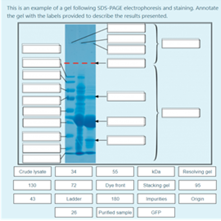
Solved This is an example of a gel following SDS PAGE | Chegg.com
InDesign Labeling / Annotating PCR Gel Pictures - YouTube In this tutorial we will learn how to annotate Agarose Gel Pictures with Adobe InDesign CS5. I see people often labeling pictures in Photoshop and I can't re...
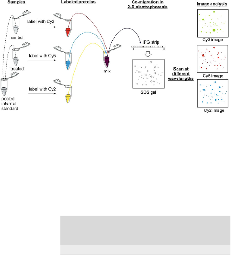
The Basics of 2D DIGE - Difference Gel Electrophoresis (DIGE ...
3 Ways to Read Gel Electrophoresis Bands - wikiHow Hold a UV light up to the gel sheet to reveal results when using a UV-based dye. With your gel sheet in front of you, find the switch on a tube of UV light to turn it on. Hold the UV light 8-16 inches (20-41 cm) away from the gel sheet. Illuminate the DNA samples with the UV light to activate the dye and read the results.

Gel Electrophoresis Products for RNa and DNA
Differential in Gel Electrophoresis - an overview - ScienceDirect After electrophoresis, the gel is scanned sequentially with the excitation wavelengths of the three dyes, and protein abundances are obtained. A given protein from different samples, regardless of the labeling dye used reaches the same position on the gel; thus problems of inter-gel variations do not interfere.
Visible Labeling of Proteins for Polyacryiamide Gel ...
PDF Lab 4: Gel Electrophoresis - Vanderbilt University Gel electrophoresis Gel electrophoresis is a method of separating DNA fragments by movement through a Jello-like substance called agarose. Derived from a seaweed polysaccharide, agarose gels form small pores that act as sieves to separate DNA based on size; whereby smaller DNA molecules move through the pores faster and easier than larger ...
Agarose gel electrophoresis of labeled DNA in which the same ...
Gel Electrophoresis - Definition, Purpose and Steps - Biology Dictionary The broad steps involved in a common DNA gel electrophoresis protocol: 1. Preparing the samples for running The DNA is isolated and preprocessed (e.g. PCR, enzymatic digestion) and made up in solution with some basic blue dye to help visualize the movement of the sample through the gel. 2. An agarose TAE gel solution is prepared

2D gel electrophoresis
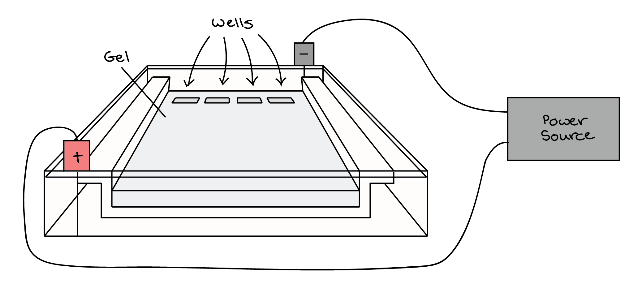
What is gel electrophoresis? - YourGenome Illustration of DNA electrophoresis equipment used to separate DNA fragments by size. A gel sits within a tank of buffer. The DNA samples are placed in wells at one end of the gel and an electrical current passed across the gel. The negatively-charged DNA moves towards the postive electrode. Image credit: Genome Research Limited.
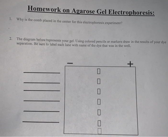
Homework on Agarose Gel Electrophoresis: 1. Why is | Chegg.com
Part 2: Analysing and Interpreting (Agarose) Gel Electrophoresis Results The agarose gel electrophoresis is a molecular genetic technique used to separate DNA on the basis of size and charge of it. The negatively charged DNA migrates towards the positive node under the influence of the current. The results of agarose electrophoresis are affected by some of the factors enlisted below, The concentration of gel

Methods in Molecular Biology: Difference Gel Electrophoresis ...
Gel Shift Assays (EMSA) | Thermo Fisher Scientific - US Gel shift assays need not be limited to protein-DNA interactions. Protein-RNA and protein-peptide interactions have also been studied using the same electrophoretic principle. Overview of the gel shift assay method. The gel shift assay consists of three key steps: (1) binding reactions, (2) electrophoresis, (3) probe detection.

Agarose Gel Electrophoresis: Results Analysis Video
Gel Electrophoresis - an overview | ScienceDirect Topics Gel electrophoresis is an analytical technique that allows size separation of DNA as well as other macromolecules. For gel electrophoresis, a DNA sample is loaded at one end of a gel matrix (usually agarose or acrylamide) that provides a uniform pore size through which the DNA molecules can move.
About 2-D Fluorescence Difference Gel Electrophoresis (2

Lab 13: Gel Electrophoresis on 5% Chelex DNA Extraction PCR ...

Synthesis of site-specific spin-labeled RNA. (A) Native gel ...

Identification of diabetes- and obesity-associated proteomic ...
01 November 2020 Todays Title CW DNA manipulation

Expression, labeling and purification. Protein expression, 35 ...

Gel electrophoresis–autoradiographic image analysis of ...
Electrophoresis

Gel electrophoresis: Pūkeko DNA — Science Learning Hub
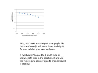
Tutorial how to make and use a standard curve gel electrophoresis

Confirmation of QD-labeling of pDNA by agarose gel ...
Molecular-weight size marker - Wikipedia
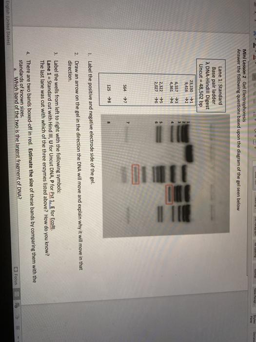
Solved Mini Lesson 2 Gel Electrophoresis Answer the | Chegg.com
Gel Electrophoresis | MMG 233 2014 Genetics & Genomics Wiki ...

PCR amplification for single-site mutagenesis. A) Agarose gel ...

Label-free Kinase Profiling Using Phosphate Affinity ...
![SDS-gel electrophoresis of [ 35 S]methionine and [ 35 S ...](https://www.researchgate.net/profile/Uddhav-Kelavkar/publication/12133784/figure/fig3/AS:341752268509188@1458491498739/SDS-gel-electrophoresis-of-35-Smethionine-and-35-Scystine-labeled-proteins-isolated.png)
SDS-gel electrophoresis of [ 35 S]methionine and [ 35 S ...
Gel electrophoresis (article) | Khan Academy

How to make a gel image using Powerpoint
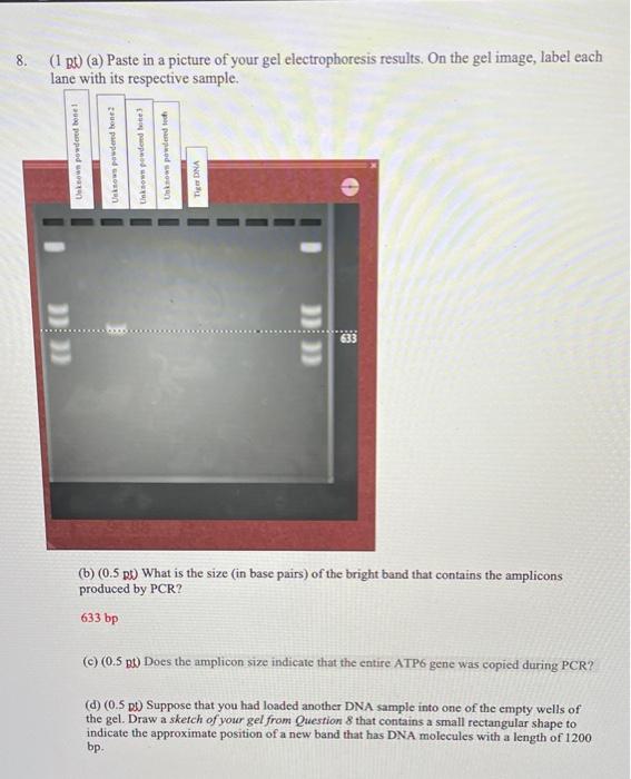
8. (1 B (a) Paste in a picture of your gel | Chegg.com

Study of Early Leaf Senescence in Arabidopsis thaliana by ...

A and B. Agarose gel electrophoresis. Lanes labelled M ...
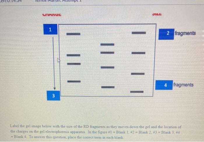
Solved UMNUE 1 2 fragments III 4 fragments 3 Label the gel ...

InDesign Labeling / Annotating PCR Gel Pictures - Advanced Tutorial (Part 12)
Gel electrophoresis (article) | Khan Academy

Internal modification and religation efficiencies. (a ...
Annotating A Gel | Get Your Science On Wiki | Fandom

Stellar Scientific Agarose LE 500g For DNA Gel Electrophoresis

Agarose gel electrophoresis of 16S-rRNA; Lane: 1-1 Kb DNA ...
Texture analysis in gel electrophoresis images using an ...

How to Interpret DNA Gel Electrophoresis Results | GoldBio
Post a Comment for "45 labeling gel electrophoresis"