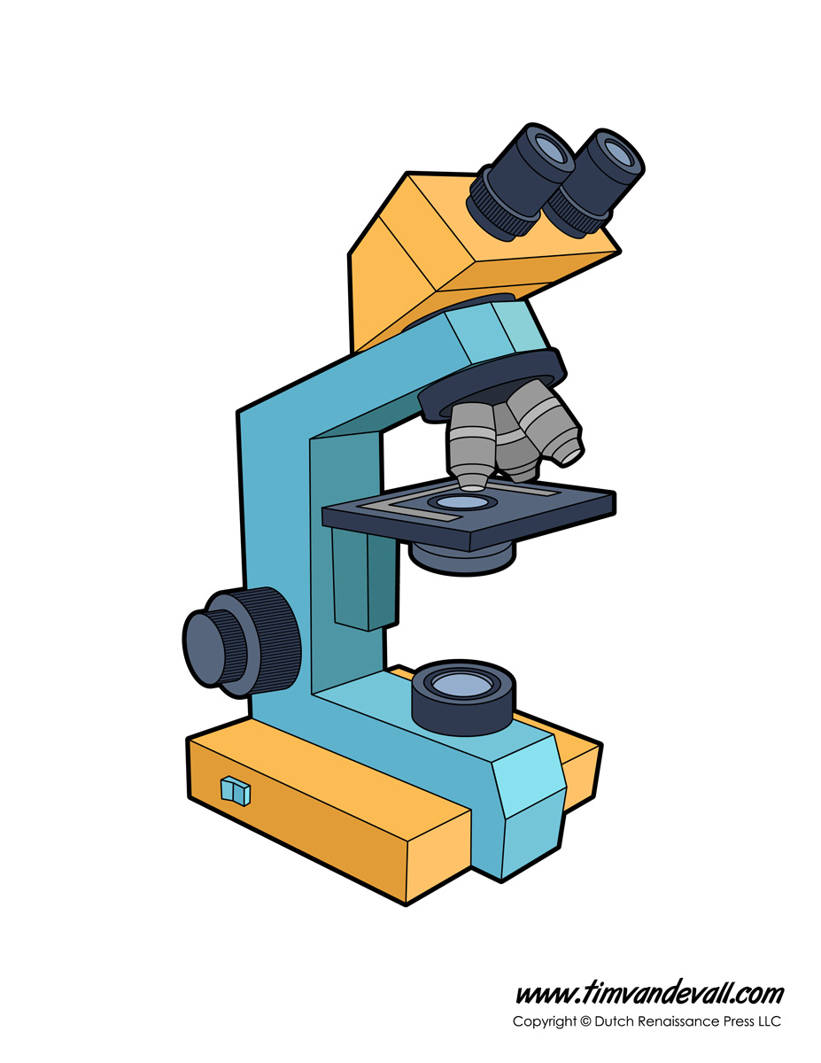
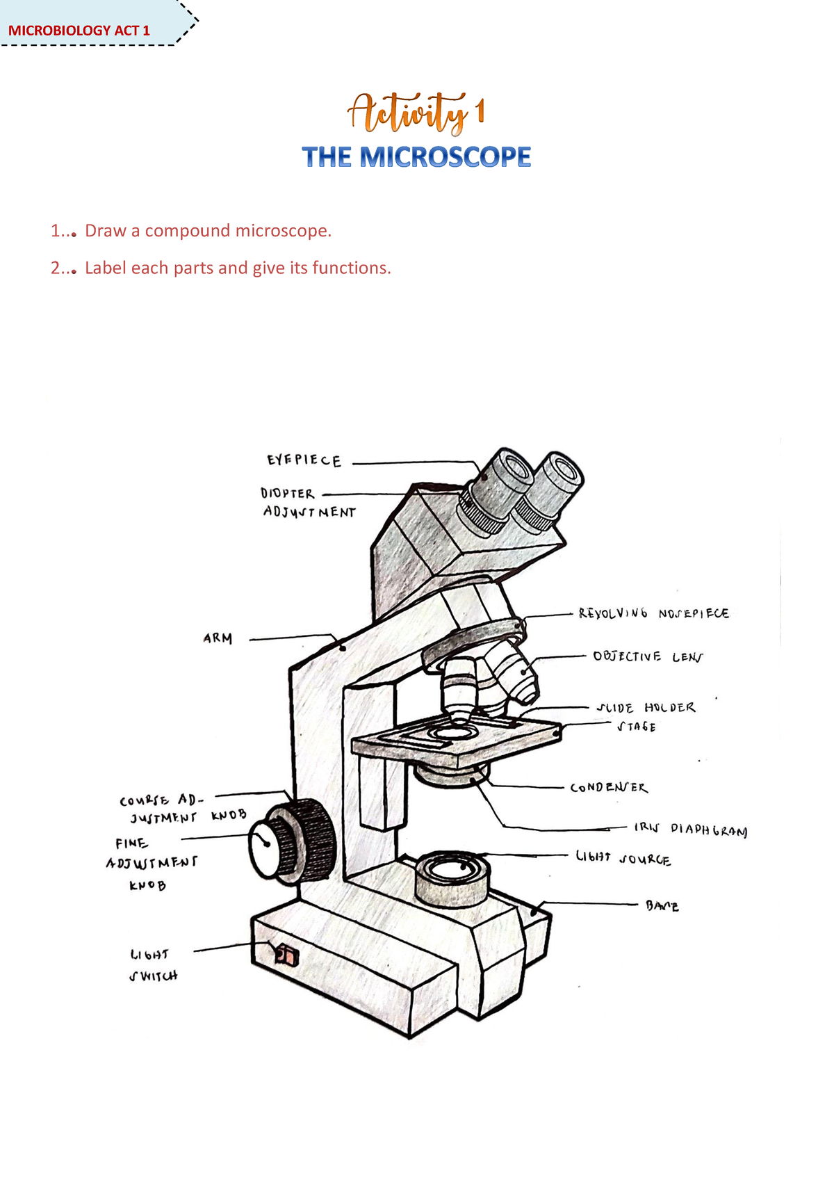
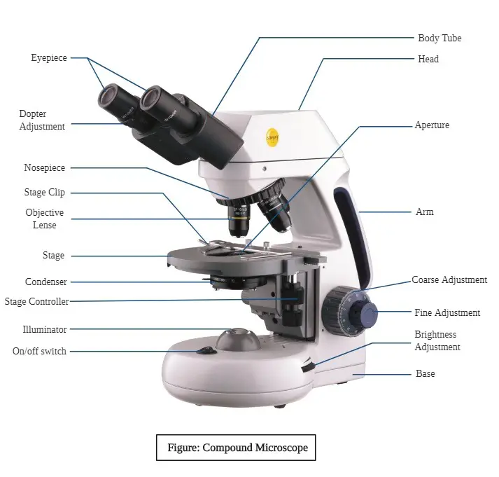
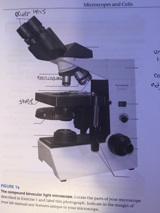
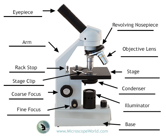
44 labeling a compound microscope
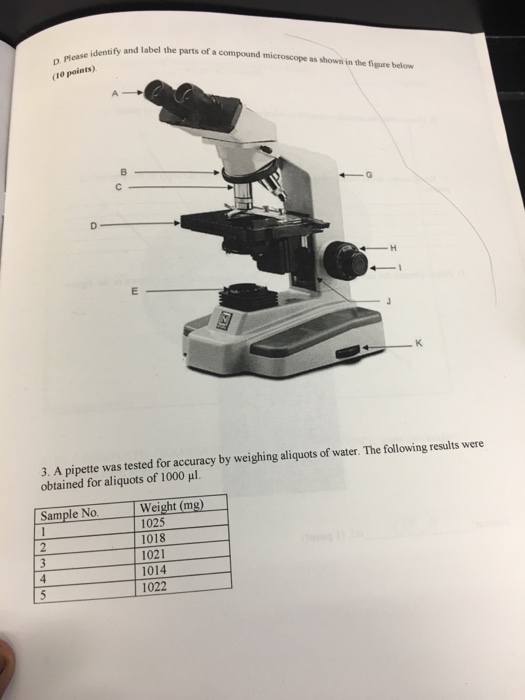
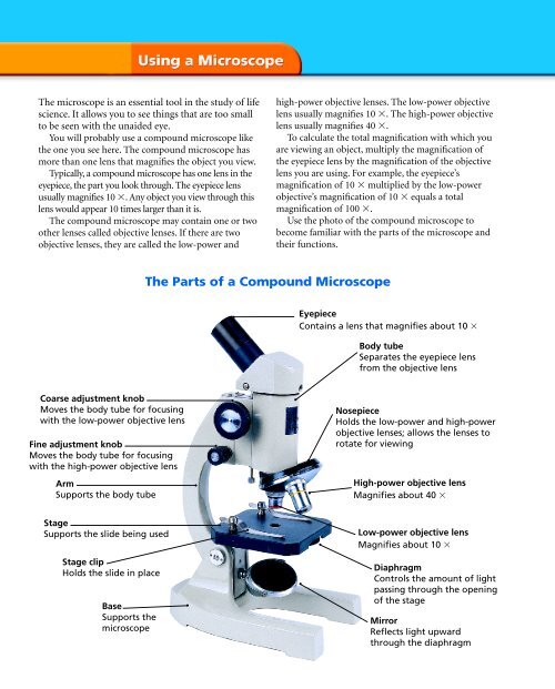
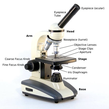
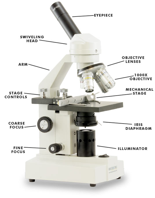
Label the parts of a microscope - AnswerData Explain why it does not belong. 7. light source, scanning power, objective lens, eyepiece 8. compound light, transmission electron, light electron, scanning electron 9. The ability of a microscope to show details clearly is called 10. The ability of a microscope to increase an object's apparent sized is called 11. label the microscope answers - Alex Becker Marketing Compound Microscope Parts - Labeled Diagram … There are three major structural parts of a compound microscope. The head includes the upper part of the microscope, which houses the most critical optical components, and the eyepiece tube of the microscope. The base acts as the … Click to visit Parts of the Microscope Labeling Activity! by Mrs …
Fundamentals Of Phlebotomy - PHS Institute Labeling the Sample 27 Equipment 27 Order of Draw 28 ... glasses, not compound microscopes of the type used today. A drawing of one of Leeuwenhoek's "microscopes" is shown at the left. Compared to modern microscopes, it ... The invention of the microscope in the 17th century, coupled with advances in

Labeling a compound microscope
Compound Microscope - Types, Parts, Diagram, Functions and Uses A compound microscope captures an inverted image of the specimen because every time the light passes through the lens, the image's direction is flipped. The image always ends up inverted from the original. So, if you move the sample to the left, it moves in the right direction. Image 18: A comparison image between a simple and compound microscope. MCQ on Microscopy Pdf - YB Study A compound microscope contains three separate lens systems. The condenser lens is placed between the light source and the specimen and it gathers and focuses the light rays in the plane of the microscopic field to view the specimen. The image formed by an objective of a compound microscope is real and enlarged. Electron Microscope- Definition, Principle, Types, Uses, Labeled Diagram There are two types of electron microscopes, with different operating styles: 1. Transmission Electron Microscope (TEM) The transmission electron microscope is used to view thin specimens through which electrons can pass generating a projection image. The TEM is analogous in many ways to the conventional (compound) light microscope.
Labeling a compound microscope. About us | Leica Microsystems Leica Microsystems develops and manufactures microscopes and scientific instruments for the analysis of microstructures and nanostructures. Widely recognized for optical precision and innovative technology, the company is one of the market leaders in compound and stereo microscopy, digital microscopy, confocal laser scanning and super-resolution microscopy with … Compound Microscope - Diagram (Parts labelled), Principle and Uses See: Labeled Diagram showing differences between compound and simple microscope parts Structural Components The three structural components include 1. Head This is the upper part of the microscope that houses the optical parts 2. Arm This part connects the head with the base and provides stability to the microscope. Brightfield Microscope (Compound Light Microscope)- Definition ... Brightfield Microscope Definition Brightfield Microscope is also known as the Compound Light Microscope. It is an optical microscope that uses light rays to produce a dark image against a bright background. It is the standard microscope that is used in Biology, Cellular Biology, and Microbiological Laboratory studies. Compound Light Microscopes | Products | Leica Microsystems 08/08/2018 · A compound microscope uses optics to produce a magnified image of a sample so that with details of it can be observed that are undetectable with the naked eye. The most basic optics of a compound microscope has at least 2 lenses: i) an objective placed nearby the sample which creates a magnified, real image of it and ii) eyepieces or oculars ...
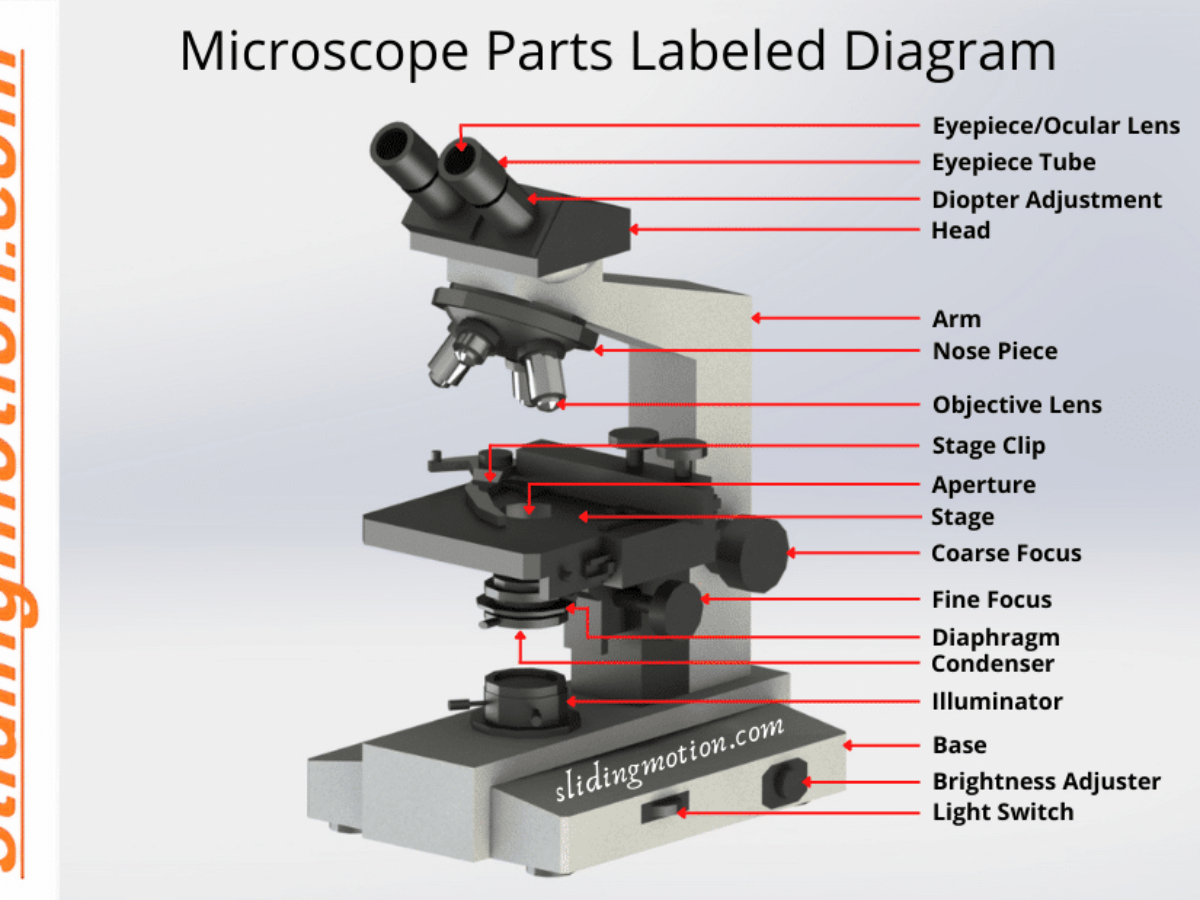
› microscope_labelingMicroscope Labeling - The Biology Corner 18. You should carry the microscope by the _____ and the _____. 19. The objectives are attached to what part of the microscope (it can be rotated to click lenses into place?) _____ 20. A microscope has an ocular objective of 10x and a high power objective of 50x, what is the microscope's total magnification? Compound Microscope Diagram Labeled - parts of a microscope ... Here are a number of highest rated Compound Microscope Diagram Labeled pictures upon internet. We identified it from trustworthy source. Its submitted by supervision in the best field. We tolerate this kind of Compound Microscope Diagram Labeled graphic could possibly be the most trending topic taking into consideration we part it in google ... › eharcourtschool-retiredeHarcourtSchool.com has been retired - Houghton Mifflin Harcourt Connected Teaching and Learning. Connected Teaching and Learning from HMH brings together on-demand professional development, students' assessment data, and relevant practice and instruction. Microscope Parts, Function, & Labeled Diagram - slidingmotion Microscope parts labeled diagram gives us all the information about its parts and their position in the microscope. Microscope Parts Labeled Diagram The principle of the Microscope gives you an exact reason to use it. It works on the 3 principles. Magnification Resolving Power Numerical Aperture. Parts of Microscope Head Base Arm Eyepiece Lens
Compound Microscope Labeling Worksheet / What Sort Of Microscopes Are ... Find label compound microscope lesson plans and teaching resources. A compound microscope is much more powerful than a simple microscope. Know the following functions of the microscope parts. Use the following list of parts of a compound microscope to label the diagram. How can a microscope help us. Here are the important compound microscope parts. › analysis › articlesAn Introduction to the Light Microscope, Light Microscopy ... Sep 02, 2021 · Figure 1: Basic compound microscope: Light from a source is focused onto the sample (object) using a mirror and condenser lens. Light from the sample is collected by an objective, forming an intermediate image which is imaged again by the eyepiece and relayed to the eye, which sees a magnified image of the sample. Microscope Parts & Specifications | Microscope World Resources The compound microscope uses lenses and light to enlarge the image and is also called an optical or light microscope (versus an electron microscope). The simplest optical microscope is the magnifying glass and is good to about ten times (10x) magnification. The compound microscope has two systems of lenses for greater magnification: 1. Simple Microscope - Parts, Functions, Diagram and Labelling A compound microscope is also called a bright field microscope. It can provide magnification by up to 1,000 times. Stereo microscope/dissecting microscope - It can magnify objects by up to 300 times. It is used to visualize opaque objects that cannot be visualized using a compound microscope.
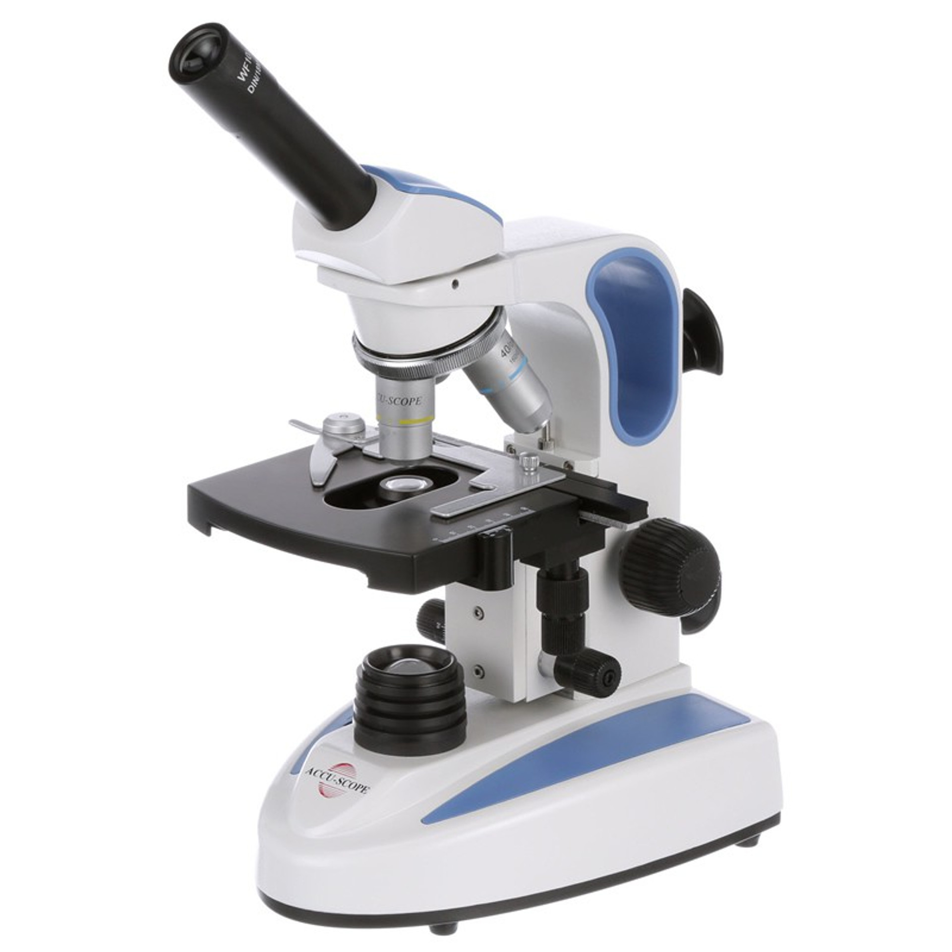
Microscope Types (with labeled diagrams) and Functions A compound microscope: Is used to view samples that are not visible to the naked eye Uses two types of lenses - Objective and ocular lenses Has a higher level of magnification - Typically up to 2000x Is used in hospitals and forensic labs by scientists, biologists and researchers to study micro organisms Compound microscope labeled diagram
eHarcourtSchool.com has been retired - Houghton Mifflin Harcourt Connected Teaching and Learning. Connected Teaching and Learning from HMH brings together on-demand professional development, students' assessment data, and relevant practice and instruction.
› light-microscopesCompound Light Microscopes | Products | Leica Microsystems Aug 08, 2018 · A compound microscope uses optics to produce a magnified image of a sample so that with details of it can be observed that are undetectable with the naked eye. The most basic optics of a compound microscope has at least 2 lenses: i) an objective placed nearby the sample which creates a magnified, real image of it and ii) eyepieces or oculars ...
› cemf › whatisemWhat is Electron Microscopy? - UMASS Medical School Conventional scanning electron microscopy depends on the emission of secondary electrons from the surface of a specimen. Because of its great depth of focus, a scanning electron microscope is the EM analog of a stereo light microscope. It provides detailed images of the surfaces of cells and whole organisms that are not possible by TEM.
Histology, Cell - StatPearls - NCBI Bookshelf 08/05/2022 · The cell is the fundamental organizational unit of life. All living things are composed of cells, which then further subdivide based on the presence or absence of the nucleus, into two types: eukaryotic cells (Greek, Eu=true, karyo=nut, nucleus) - these cells are present in all the human, animal and plants with a clear, distinct nucleus. Prokaryotic cells are some bacteria …
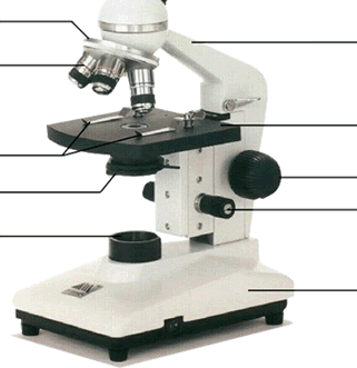
Parts of a microscope with functions and labeled diagram - Microbe Notes Q. List down the 18 parts of a Microscope. 1. Ocular Lens (Eye Piece) 2. Diopter Adjustment 3. Head 4. Nose Piece 5. Objective Lens 6. Arm (Carrying Handle) 7. Mechanical Stage 8. Stage Clip 9. Aperture 10. Diaphragm 11. Condenser 12. Coarse Adjustment 13. Fine Adjustment 14. Illuminator (Light Source) 15. Stage Controls 16. Base 17.
Compound Microscope- Definition, Labeled Diagram, Principle, Parts, Uses Alternatively, the magnification of the compound microscope is given by: m = D/ fo * L/fe where, D = Least distance of distinct vision (25 cm) L = Length of the microscope tube fo = Focal length of the objective lens fe = Focal length of the eye-piece lens Parts of a Compound Microscope Eyepiece And Body Tube.
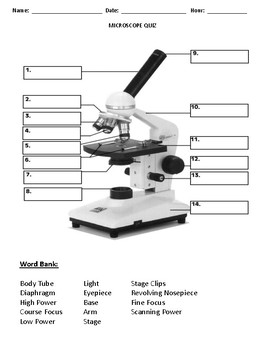
Microscope Labeling - The Biology Corner Students label the parts of the microscope in this photo of a basic laboratory light microscope. Can be used for practice or as a quiz. Name_____ Microscope Labeling . Microscope Use: 15. When focusing a specimen, you should always start with the _____ objective. 16. When using the high power objective, only the _____ knob should be used.
What is Electron Microscopy? - UMASS Medical School Electron microscopy is used in conjunction with a variety of ancillary techniques (e.g. thin sectioning, immuno-labeling, negative staining) to answer specific questions. ... (compound) light microscope. TEM is used, among other things, to image the interior of cells (in thin sections), the structure of protein molecules (contrasted by metal ...
Microscope parts/labeling Label the image of a compound light ... Microscope parts/labeling Label the image of a compound light microscope using the terms provided. Questions & Answers. accounting; computer-science ... Using the following list, label the parts of the bright-field compound microscope: base, dialirheostat, iris diaphragm, light souree, mechanical stage knobs, objeetive, ocular, pinion knob ...
› companyAbout us | Leica Microsystems Leica Microsystems develops and manufactures microscopes and scientific instruments for the analysis of microstructures and nanostructures. Widely recognized for optical precision and innovative technology, the company is one of the market leaders in compound and stereo microscopy, digital microscopy, confocal laser scanning and super-resolution microscopy with related imaging systems ...
An Introduction to the Light Microscope, Light Microscopy Techniques ... 02/09/2021 · Figure 1: Basic compound microscope: Light from a source is focused onto the sample (object) using a mirror and condenser lens. Light from the sample is collected by an objective, forming an intermediate image which is imaged again by the eyepiece and relayed to the eye, which sees a magnified image of the sample.
Electron Microscope Ans: A light microscope has a low resolving power (0.25µm to 0.3µm) while the electron microscope has a resolution power about 250 times higher than the light microscope at about 0.001µm. Similarly, a light microscope has a magnification of 500X to 1500x while the electron microscope has a much higher magnification of 100,000X to 300,000X.
US10379038B2 - Measuring a size distribution of nucleic acid … A process for measuring a size distribution of a plurality of nucleic acid molecules, the process comprising: labeling the nucleic acid molecules with a fluorescent dye comprising a plurality of fluorescent dye molecules to form labeled nucleic acid molecules, such that a number of fluorescent dyes molecules attached to each nucleic acid molecule is reliably proportional to …
Simple Microscope - Diagram (Parts labelled), Principle, Formula and Uses The magnification power of a simple microscope is expressed as: M = 1 + D/F Where M = Magnification power D = the lease possible distance of distinct vision of eye, typically 25cm F = Focal length of the convex lens It is to be noted that
› t-partsMicroscope Parts & Specifications | Microscope World Resources The compound microscope uses lenses and light to enlarge the image and is also called an optical or light microscope (versus an electron microscope). The simplest optical microscope is the magnifying glass and is good to about ten times (10x) magnification. The compound microscope has two systems of lenses for greater magnification: 1.
Parts of the Microscope with Labeling (also Free Printouts) Parts of the Microscope with Labeling (also Free Printouts) By Editorial Team March 7, 2022 A microscope is one of the invaluable tools in the laboratory setting. It is used to observe things that cannot be seen by the naked eye. Table of Contents 1. Eyepiece 2. Body tube/Head 3. Turret/Nose piece 4. Objective lenses 5. Knobs (fine and coarse) 6.
compound light microscope labeled Labeling the Parts of the Microscope. Anand's Human Anatomy for Dental Students, Third Edition - Page 488 Brightfield Light Microscope (Compound light microscope) This is the most basic optical With contributions by numerous experts Fundamentals of Light Microscopy and Electronic Imaging Eyepiece (ocular lens) with or without Pointer: The part ...
Microscope Quiz: How Much You Know About Microscope Parts ... - ProProfs The microscope has been used in science to understand elements, diseases, and cells. You must have used a microscope back in high school in the biology lab. Do you believe you understood how to use it? Take up the test and see. ... Parts Of The Compound Light Microscope Quiz. This should help you study the functions of a microscope for the ...
Electron Microscope- Definition, Principle, Types, Uses, Labeled Diagram There are two types of electron microscopes, with different operating styles: 1. Transmission Electron Microscope (TEM) The transmission electron microscope is used to view thin specimens through which electrons can pass generating a projection image. The TEM is analogous in many ways to the conventional (compound) light microscope.
MCQ on Microscopy Pdf - YB Study A compound microscope contains three separate lens systems. The condenser lens is placed between the light source and the specimen and it gathers and focuses the light rays in the plane of the microscopic field to view the specimen. The image formed by an objective of a compound microscope is real and enlarged.
Compound Microscope - Types, Parts, Diagram, Functions and Uses A compound microscope captures an inverted image of the specimen because every time the light passes through the lens, the image's direction is flipped. The image always ends up inverted from the original. So, if you move the sample to the left, it moves in the right direction. Image 18: A comparison image between a simple and compound microscope.






(159).jpg)








Post a Comment for "44 labeling a compound microscope"